产品简介:
DiSC3(5) (3,3'-Dipropylthiadicarbocyanine Iodide)
重要提醒(购买或初次使用前)请务必查阅:
|
一、荧光染料(粉末形式,特别是对氧敏感探针)配制储存液的注意事项
|
|
1)荧光探针在固体(粉末)状态很稳定,按照说明书要求温度来保存,效期内使用即可。
2)荧光探针用有机溶剂比如:DMSO,溶解配制成储存液之后,一般来说,放到-20℃保存(2-3个月内使用,甚至更短,具体可咨询);放到-80℃保存(6个月内使用,具体可咨询)。前提是,使用的有机溶剂必须是高质量且无水的,特别是DMSO,必须是新鲜开封。
3)配制的储存液请务必用密封性好且螺旋盖的低容量冻存管保存(不可用EP管),至少按照5-10ul/管来分装,避光冻存。
4)对于某些特殊化合物(对空气敏感或存在不稳定结构),可能保存周期特别短,甚至只能当天使用。这些化合物说明书上会有说明。
有更多信息,请联系我司工作人员来核实。
|
|
二、荧光染料(以溶于有机溶剂的储存液形式提供)的注意事项
|
|
1) 以溶于有机溶剂的储存液形式的荧光探针相对来说是比较稳定的化合物;但收到这类产品,也需用户根据单次用量(5-10ul/管来分装),-20℃以下密封避光保存,减少反复冻融次数。
2) 请务必用密封性好且螺旋盖的低容量冻存管保存(不可用EP管)。务必避光。
有更多信息,请联系我司工作人员来核实。
|
关键词:
DiSC3(5);DiOC5(3);DiOC6(3);ER-Tracker Blue-White DPX;Membrane depolarization assay膜去极化实验;CAS:53213-94-8;
订购信息:
|
产品名称
|
产品编号
|
规格
|
价格(元)
|
DiSC3(5) (3,3'-Dipropylthiadicarbocyanine Iodide)
|
MX4033-10MG
|
10mg
|
498
|
|
DiSC3(5) (3,3'-Dipropylthiadicarbocyanine Iodide)
|
MX4033-50MG
|
50mg
|
998
|
|
DiSC3(5) (3,3'-Dipropylthiadicarbocyanine Iodide)
|
MX4033-250MG
|
250mg
|
2148
|
产品描述
DiSC3(5) (3,3'-Dipropylthiadicarbocyanine Iodide),一种具短(C3)烷基尾的羰花青染料,这种阳离子染料能用来检测和测量由膜改性试剂引起的跨膜电位或结构变化。
产品特性
-
CAS NO:53213-94-8
-
化学名:Benzothiazolium, 3-propyl-2-(5-(3-propyl)-2(3H)-benzothiazolidene-1,3-pentadienyl), iodide
-
英文同义名:diS-C3-(5); 3,3'-Di-n-propylthiadicarbocyanine iodide; 3,3-Dipropylthiadicarbocyanine iodide; 5-(3-Propylbenzothiazol-2-ylidene)-1-(3-propylbenzothiazolium-2-yl)-1,3-pentadiene Iodide; 3-Propyl-2-[5-[3-propyl-2(3H)-benzothiazolylidene]-1,3-pentadienyl]benzothiazolium iodide;
-
中文同义名:3,3'-二丙基硫杂二羰花青碘化物;碘化-3,3ˊ-二丙基硫杂二羰花青;
-
分子式:C25H27IN2S2
-
分子量:546.53g/mol
-
纯度:≥98%(HPLC)
-
Ex/Em:500/705 nm (in DMF)
-
溶解性:溶于DMF
-
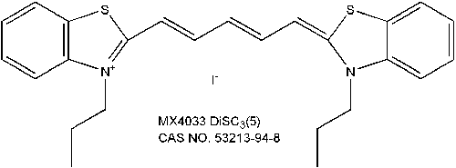
化学结构式:
保存与运输方法
保存:室温避光干燥保存,可置于-20℃长期干燥避光保存,2年有效。
运输:室温运输。
应用示例:
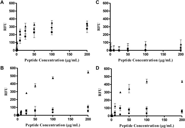
文献来源:Juba ML et al. Helical cationic antimicrobial peptide length and its impact on membrane disruption. Biochim Biophys Acta. 2015 May;1848(5):1081-91. PMID: 25660753
实验方法(膜去极化实验MK):Frozen aliquots ofenumerated bacteria (E. coli or B. cereus)were thawed on ice and washed 3 times with buffer (5 mM HEPES with 20 mM glucose, pH 7.4). Following washing, the pelleted bacteria were re-suspended in HEPES buffer (5 mM HEPES, pH 7.4, 20 mM glucose) containing either, 10 or 100 mM KCl. A 96-well plate was prepared where wells are charged with360 μL of bacterial suspension (2 × 10⁷ CFU/mL) and 4.19 μL of diSC3-(5) (200 nM) for a total volume of 364.19 μL.The bacteria were incubated at room temperature and fluorescence was monitored (622 nmex/670 nmem) until diSC3-(5) maximal uptake was obtained. Maximal diSC3-(5) uptake is indicated by a baseline in fluorescence due to self-quenching as the dye concentrates in the cell membrane. Peptide (NA-CATH, L- or D-ATRA-1A) was added at varied concentrations (200–2 μg/mL) in 20 μL aliquots and the fluorescence increase due to induced depolarization of the cytoplasmic membrane was recorded. A negative control of bacteria and diSC3-(5) was used as a background. As a positive control, complete collapse of the membrane potential was attained with valinomycin (200–2 μg/mL), a potassium ionophore.
检测原理(MK-DiSC3-(5)的膜去极化实验):DiSC3(5)作为一种膜电位敏感探针,聚集在磷脂双分子层内,引起染料的自淬灭。当膜改性化合物(比如多肽)使膜去极化,电位丧失,DiSC3(5)释放进入溶液引起荧光增强,荧光强弱与电位减少程度呈正比。
结果展现(MK-DiSC3-(5)的膜去极化实验):
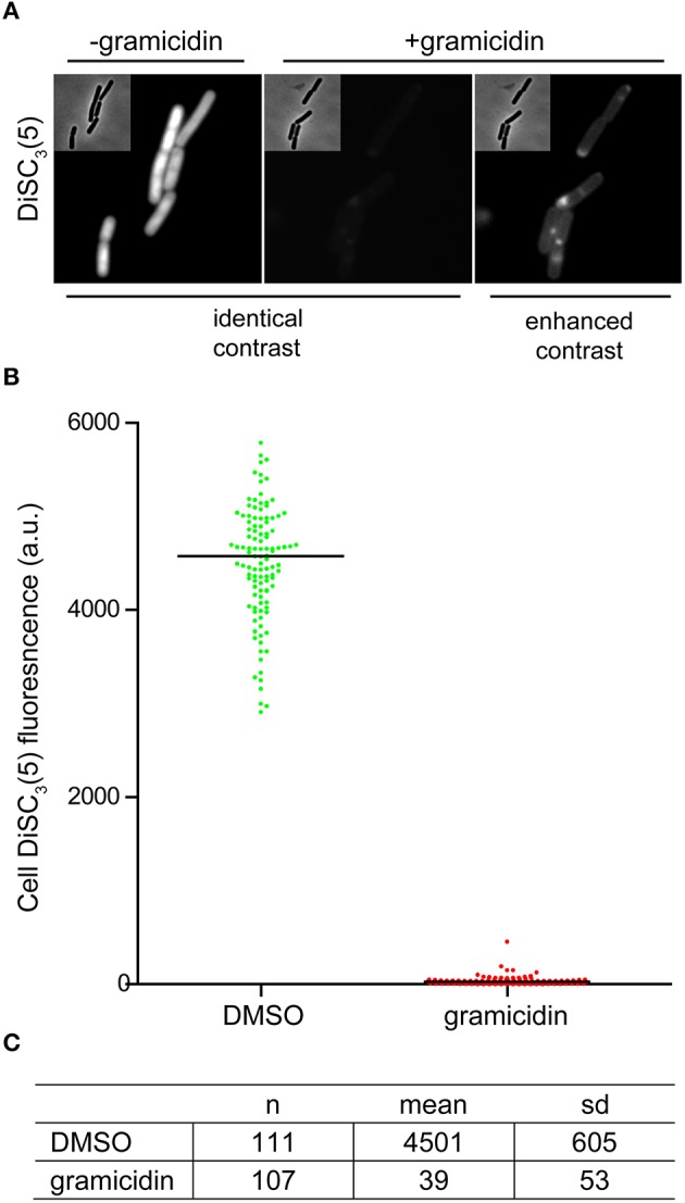
文献来源:Te Winkel JDet al.Analysis of Antimicrobial-Triggered Membrane Depolarization Using Voltage Sensitive Dyes. Front Cell Dev Biol. 2016 Apr 13;4:29. PMID: 27148531
实验方法(膜电位的显微分析MK):Early-mid logarithmic growth phase cell suspensions were incubated with 2 μM DiSC3(5)directly in the growth medium. The incubation was carried out under shaking for 5 min, immediately followed by microscopy.A final concentration of 0.5–1% DiSC3(5)-solvent dimethyl sulfoxide (DMSO) was found to be crucial in order to maintain appropriate solubility;lower solvent concentrations resulted in a strongly reduced cellular fluorescence. The incubation was carried out at growth temperature, and under vigorous shaking in order to maintain good energization of the cells. This step was routinely carried out with 2 ml round bottom Eppendorf tubes containing 200 μl cell suspensionin a thermomixer. To provide sufficient aeration, the lids of the tubes were perforated. When dissipation of membrane potential was tested, a compound of interest was added in parallel to DiSC3(5).Addition of 5 μM gramicidin (a mixture of gramicidin A, B, C, and D)was routinely used as a positive control. The imaging of DiSC3(5)-stained cells was carried out using commonly availableCy5-filter sets.【重要信息:文章多次提及到DiSC3(5)的工作浓度内维持1%DMSO浓度,对维持染料的溶解性和荧光稳定性很是关键。】
结果展现(MK-DiSC3-(5)的单细胞膜电位显微分析):
注意事项
1)荧光染料均存在淬灭问题,请尽量注意避光,以减缓荧光淬灭。
2) 为了您的安全和健康,请穿实验服并戴一次性手套操作。
相关产品:
|
货号
|
名称
|
规格
|
|
MX4008-100MG
|
DiOC2(3)绿色膜电位荧光探针
|
100mg
|
|
MX4009-10MG
|
DiOC6(3)内质网荧光探针
|
10mg
|
|
MX4032-25MG
|
DiOC5(3)膜电位荧光探针
|
25mg
|
|
MX4033-50MG
|
DiSC3(5) (3,3'-Dipropylthiadicarbocyanine Iodide)
|
50mg
|
|
MS0049-5MG
|
Valinomycin缬氨霉素
|
5mg
|
|
MS0078-50MG
|
Gramicidin短杆菌肽
|
50mg
|
|
MX4351-50UL
|
ER-Tracker Blue-White DPX, for live-cell imaging
|
50µl
|
|
MX4352-20UL
|
ER-Tracker Green (BODIPY™ FL Glibenclamide), for live-cell imaging
|
20µl
|
|
MX4353-20UL
|
ER-Tracker Red (BODIPY™ TR Glibenclamide), for live-cell imaging
|
20µl
|
——Written/Edited by V. Shallan【版权归MKBio懋康所有】
上海懋康生物科技有限公司是一家涉足于生命科学和生物技术领域研究的试剂、仪器和实验室消耗品与实验服务工作,主要从事细胞生物学、植物学、分子生物学、免疫学、生物化学、蛋白组学。生物制药与诊断试剂研发生产等领域。 本公司秉承“以人为本,以诚为信、合同守信”的经营理念。坚持"品质保障"的原则为广大客户提供优质产品。
引用文献:
[1]Zhu YY, Wang ZJ, Zhu M, Zhou ZS, Hu BY, Wei MZ, Zhao YL, Dai Z, Luo XD. A dual mechanism with H2S inhibition and membrane damage of morusin from Morus alba Linn. against MDR-MRSA. Bioorg Med Chem. 2024 Jan 1;97:117544. doi: 10.1016/j.bmc.2023.117544. Epub 2023 Dec 6. PMID: 38071943.
SYTOX Green nucleic acid stain and 3,3′-dipropylthiadicarbocyanine iodide (DiSC3(5)) were purchased from Shanghai Maokang Biotechnology Co., Ltd. (Shanghai, China).
[2]Duan Y, Wang ZJ, Mei LN, Shen JS, He XC, Luo XD. Anti-Candida albicans effect and mechanism of Pachysandra axillaris Franch. J Ethnopharmacol. 2025 Jan 31;340:119284. doi: 10.1016/j.jep.2024.119284. Epub 2024 Dec 24. PMID: 39725364.
3,3′-dipropylthiadicarbocyanine iodides [DiSC3 (5)] (Maokang Biotechnology Co., Ltd. Shanghai, China) was used to evaluate the membrane depolarization state of fungi
[3] Huang K, Liu W, Zhao FJ. Methylarsenite is a broad-spectrum antibiotic disrupting cell wall biosynthesis and cell membrane potential. Environ Microbiol. 2023 Feb;25(2):562-574. doi: 10.1111/1462-2920.16309. Epub 2022 Dec 19. PMID: 36510854.
t he 0.5 μl of 3 mM DISC 3 -(5) (Mkbio, China) dissolved in dimethyl sulfoxide (DMSO)
was added to each of the cell suspensions and mixed thoroughly.
|


 沪公网安备31011202021859号